

Master pathologist offers a primer on prostate biopsy
Part 1.

(Note: Dr. Ming Zhou’s column provides a short course on prostate pathology for patients with prostate cancer. His “The Pathology Report” column demystifies the prostate biopsy pathology reports to help patients like you and me understand how the pathology results may influence prostate cancer management decisions. At a reader’s request, Ming here is providing a primer on biopsies for those of us who get them.—Howard Wolinsky)
How Prostate Biopsy Specimens Are Processed: From Patients to Pathologists (Part 1)
Prostate biopsy is the gold standard for diagnosing prostate cancer. This article provides an overview of how prostate biopsies are performed, processed in pathology laboratories, and reviewed by pathologists. Understanding this process can help you appreciate the pros and cons, including the limitations, of the prostate biopsy. This discussion is divided into two parts, with Part 1 focusing on the biopsy procedure and initial specimen processing. (Part 2 will appear in a June issue of TheActiveSurveillor.com.)
1. How Prostate Biopsy Is Done by Urologists
Prostate biopsy differs significantly from biopsies of other organs, such as the lung or breast, where only lesions visible on imaging are biopsied. In the prostate, cancerous lesions may not be reliably detected by imaging, and often there are multiple samples. Therefore, a systematic biopsy, also known as the sextant biopsy, is used. This involves obtaining tissue samples (prostate cores) from six predetermined areas of the prostate gland: medial and lateral regions of the apex, mid-gland, and base on both sides.
Prostate biopsies can be performed using either a transrectal or transperineal approach, each with its advantages and disadvantages (Reference 1).
A. Transrectal Approach
Procedure: A biopsy needle is inserted with ultrasound guidance through the rectum into the prostate to sample tissue from the six predefined regions.
Advantages: This technique is more familiar to urologists.
Disadvantages: It may pose a higher risk of infection, including urinary tract infections and, more rarely, serious complications such as sepsis. It may also inadequately sample certain regions of the prostate, particularly the anterior and apical regions.
B. Transperineal Approach
Procedure: The needle is inserted through the skin between the scrotum and anus.
Advantages: Lower risk of infection and better access to the anterior region of the prostate, reducing the risk of missed cancers.
Disadvantages: Technically more challenging and requires specialized equipment and expertise.
Recently, MRI-guided prostate biopsy, which targets suspicious areas identified on multiparametric magnetic resonance imaging (mpMRI), has been increasingly used in conjunction with systematic biopsies. This method can increase the likelihood of detecting clinically significant prostate cancer while reducing the detection of insignificant or low-grade cancers.
2. Size of Needle Cores
Typically, one tissue core is taken from each of the six predetermined regions of the prostate gland, plus 3-6 cores from MRI-identified abnormal regions during MRI-targeted biopsy. The tissue cores obtained are typically 18 gauge (0.87 mm in diameter) and approximately 12 millimeters in length.

(Two prostate cores, shown here as 1.0- and 1.5-cm long thin tissue threads.)
3. Processing of Prostate Biopsy Specimens in Pathology Laboratories
When prostate biopsy specimens arrive in pathology labs, they undergo several steps before being examined under a microscope by pathologists. Each step is crucial and can affect the quality of the diagnosis.
A. Fixation: After collection, biopsy cores are immediately placed in fixative solutions, commonly formalin, to prevent degradation and preserve the tissue structure for accurate histological assessment and potential molecular studies.

(Prostate biopsy cores are immediately put in containers with formalin for fixation.)
B. Microtomy: Fixed tissue cores are processed by embedding them in paraffin wax to harden the tissue, facilitating thin sectioning. A microtome is used to cut thin slices (typically 4-5 micrometers thick) from the embedded tissue blocks. These slices are mounted onto glass slides for staining.

(Prostate cores (pink tissue thread) are embedded in paraffin wax to form tissue blocks.)

(Tissue blocks containing prostate cores are sliced on a microtome into thin slices, typically 4-5 micrometers thick, and mounted on glass slides.)
C. Staining: The glass slides with mounted tissue sections are stained with dyes (hematoxylin and eosin [H&E]) to highlight cellular structures, allowing pathologists to visualize the tissue morphology and architecture. Additional stains, such as immunohistochemistry, may be used to detect basal cells absent in cancer glands.

(A prostate biopsy glass slide with 2 tissue sections.)
D. Amount of Tissue on Glass Slide: Multiple tissue sections from each biopsy core are mounted onto individual glass slides. Pathologists examine several slides to ensure a comprehensive evaluation of the entire biopsy specimen.

(An H&E slide of prostate needle biopsy with multiple tissue cores.)
4. Sampling Limitations of Prostate Biopsy
Despite careful biopsy techniques, sampling errors can occur due to the small size of the biopsy cores and the heterogeneous nature of prostate cancer. Consider the following facts:
Prostate Size: A normal prostate is approximately 3x3x5 cm, or 25 mL.
Biopsy Volume: An 18 gauge needle biopsy generates about 12 mm-long, 0.838 mm- thick cylindrical tissue with a volume of 0.007 mL, 0.028% of a prostate gland. It will take roughly 5,000 cores to biopsy the entire prostate gland. If 12 cores are taken, that’s about 0.3% of the prostate gland being biopsied.
Microscopic Examination: Pathologists only examine around 5% of the biopsy tissue on slides.
Overall Sampling: Ultimately, pathologists review about 0.015%, or roughly 0.02%, of the prostate gland.
This means that prostate biopsies sample a tiny fraction of the prostate gland, potentially missing areas of cancer or the worst areas of cancer, leading to false-negative results or undergrading (the biopsy grade being falsely lower than the actual cancer grade in the prostate). Conversely, overgrading can also occur.

(A comparison of prostate gland (roughly 3x3x5 cm) and 2 prostate needle cores. Volume-wise, a set of prostate needle cores account for <0.3% of the prostate gland.)
Doctors and patients should be aware of the limitations of biopsy sampling and correlate histological findings with clinical and imaging data to minimize diagnostic errors. Despite these limitations, prostate biopsy is remarkably effective in predicting clinical outcomes. Higher Gleason scores (grade groups) and larger tumor volumes in prostate biopsy are generally associated with poorer clinical outcomes, including higher rates of cancer progression, metastasis, and mortality.
Reference:
Chung Y, Hong SK. Shifting to transperineal prostate biopsy: A narrative review. Prostate Int. 2024 Mar;12(1):10-14. doi: 10.1016/j.prnil.2023.11.003. PMID: 38523899
Dr. Zhou is the Chair and Pathologist-in-Chief of the Tufts Medical Center, and Professor and Chair of the Department of Anatomic and Clinical Pathology, Tufts University School of Medicine in Boston. He has published over 200 peer-reviewed articles and numerous book chapters and edited five textbooks of urological and prostate pathology. He is currently a member of the United States and Canadian Academy of Pathology Board of Directors, and the immediate past President of the Genitourinary Pathology Society (GUPS), an international organization for urological pathologists.
Please send questions to mailto:pros8canswers@gmail.com or cut and paste: pros8canswers@gmail.com
Keep the questions short and sweet. They should be of general interest. Sign with your real name, or just initials, tell me where you live, how long you‘ve been on AS, how it’s going for for you. Share a whimsical signature if you’re so inclined.
Please come and invite your sons to ASPI webinar on May 25—Prostate cancer not just a country for old men.
By Howard Wolinsky
Prostate cancer typically is diagnosed in men in their late 60s. It’s considered a disease of aging, an old man’s disease.
But Gabe Canales tells a different story.
He was diagnosed when he was 35. At an upcoming webinar for Active Surveillance Patients International (ASPI), Canales will share his prostate cancer experience and how it can help you and your sons and grandsons.
The webinar will be at noon Eastern Saturday, May 25. Register Here: https://tinyurl.com/3sexhrrp
Mark Lichty, chairman of ASPI, said: “This is a session that men and their sons should attend. I urge men with prostate cancer to invite their sons.”
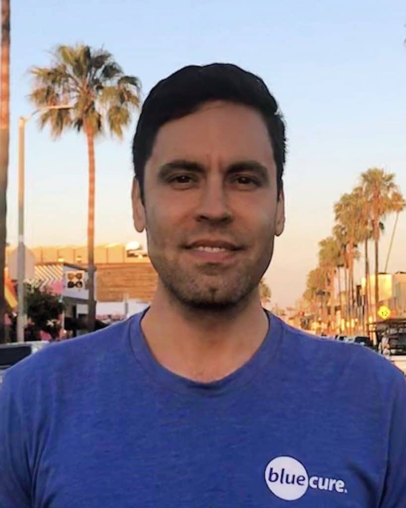
(Gabe Canales, of Blue Cure, https://bluecure.org/)
6-time Grammy-winning saxophonist David Sanborn dies from PCa
By Howard Wolinsky
David Sanborn, the influential saxophonist whose Grammy-winning career included collaborations with Stevie Wonder, Paul Simon, and David Bowie, died May 12 from advanced prostate cancer. He was 78.

“Mr. Sanborn had been dealing with prostate cancer since 2018, but had been able to maintain his normal schedule of concerts until just recently,” his team said in an X (formerly Twitter) thread. “Indeed he already had concerts scheduled into 2025.”
Sanborn collaborated with an impressive list of elite musicians, including Stevie Wonder, the Rolling Stones, David Bowie, Paul Simon, James Taylor, Luther Vandross, and Eric Clapton, according to his website.
“I sometimes get looped in with jazz musicians because I play sax and improvise,” Sanborn told The LA Times in 1996. “But if you know my music, you wouldn’t confuse it with jazz. There are certain stylistic and rhythmic elements that keep me from being in that category.”
Among pop and R&B fans, Sanborn is best known for his work on Wonder’s “Talking Book,” Bowie’s “Young Americans” and records by Bonnie Raitt and Chaka Khan.
Sanborn earned 16 Grammy Award nominations and six wins, including honors for his albums “Straight to the Heart” and “Double Vision.” Sanborn released 25 albums, including eight that went gold and one that achieved platinum status.
“David Sanborn was a seminal figure in contemporary pop and jazz music,” Monday’s statements added. “It has been said that he ‘put the saxophone back into Rock ’n Roll.’”
Unsolicited testimonial from a subscriber
"You seek out the truth of the science - just like a real journalist should."
Great information on biopsies, Howard. Thanks. It’s certainly not an exact science, as we found out after having samples read by 4 different pathologists at 4 different institutions before 2 reports agreed.
On young men affected, metastatic prostate cancer claimed my brother’s stepson, a wonderful young man, at age 36. Please read: Jeremy Brandon Paster
https://www.eastbaytimes.com/obituaries/jeremy-brandon-paster/
He wasn’t diagnosed until he was suffering from back pain from all the mets. His mom became a board member of Zero after his death to advocate for earlier routine testing.
This is yet another greatly helpful expertly-written publication from you, Howard. Ming's insights and knowledge have been a tremendous blessing to Keith Day and myself. Thank you for The Active Surveillor and the encouragement it has provided us both as well so many other men!